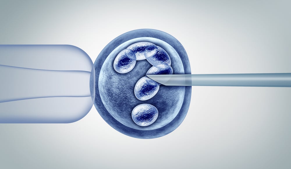
The IVF Process: How Does it Work?

For many couples suffering with infertility, in vitro fertilization (IVF) is the most effective, and often the only treatment, that will allow for conception to occur. In cases in which a woman’s fallopian tubes are damaged or removed, or there is a severe sperm abnormality, or when attempts to conceive using more conservative measures fail, IVF is quite likely to be highly successful. IVF involves the removal of a woman’s eggs from her ovaries and fertilization of the eggs with the sperm of her partner (or donor) in a specialized laboratory. The IVF process begins with daily injections of the same hormones the brain makes to stimulate the ovaries during a woman’s natural menstrual cycle. In IVF, these hormone injections are typically administered for 9 to 11 days, during which time the fertility doctor intermittently monitors the growth of the follicles (structures within the ovaries that contain the eggs) using vaginal sonograms and blood hormone testing. Once the follicles have grown to an optimal size, the woman undergoes intravenous anesthesia for 10-15 minutes and a needle is passed through the walls of the vagina into the ovaries to retrieve her eggs. As the egg retrieval is being performed, the male partner produces a semen sample (or a frozen sperm sample is thawed) and the sperm is isolated.
After the egg retrieval, the couple (or woman) returns home and the sperm is cultured together with the eggs in an incubator, or in some cases injected into the eggs (intracytoplasmic sperm injection, or ICSI). The next morning, the embryology team checks to see how many eggs have fertilized (now called embryos). The embryos are cultured in the laboratory for 5-7 days and either transferred into the uterus in the same treatment cycle after additional hormone preparation of the endometrium (inner lining of the uterus), or biopsied to obtain cells that will be sent for chromosome testing to determine if the embryos have a normal number of chromosomes (preimplantation genetic testing, or PGT). In the latter case, the embryos are cryopreserved (frozen) until the chromosome results are completed (typically within 7 days), and then a normal embryo will be selected, thawed, and transferred into the uterus after 10 -14 days of hormone preparation designed to optimize the synchrony between the endometrium and the embryo required for a successful pregnancy. A pregnancy test is performed nine days after the embryo transfer.
Extraordinary technological improvements in genetic testing of embryos have allowed reproductive endocrinologists to select the embryo most likely to implant in the uterus and lead to a normal pregnancy. At times, however, the transfer of a chromosomally normal embryo fails to result in successful implantation and pregnancy. Continuous research is being done to better understand the process of implantation during which an embryo adheres to, and invades into, the wall of the uterus to gain a rich supply of blood, oxygen and nutrients from the mother. In natural pregnancies, implantation of the fertilized egg (blastocyst) typically occurs 6 to 10 days after ovulation. The four day time frame optimal for implantation is known as the “implantation window.”
In preparation for implantation, the blood supply to the inner lining of the uterus (endometrium) increases from the time of ovulation and to its peak approximately seven days after ovulation. During this time, levels of certain hormones, particularly progesterone, increase within the endometrium while levels of other hormones decrease. Specialized cells known as decidual cells begin to cover the inner surface of the uterus and endometrial glands begin to secrete nutrients (e.g lipids and glycogen) and various hormones and proteins that allow the embryo to implant and grow. At the same time, the blastocyst sheds the thick membrane (zona pellucida) that protected it during its travel to the uterus, and the embryo attaches to and invades the uterine wall. As the pregnancy progresses, the placenta is formed, and it becomes the organ through which nutrients are passed from mother to fetus and waste materials from fetus to mother.
A fascinating and still not well-understood question is why the mother’s immune system does not reject the embryo and automatically expel or destroy it. After all, the embryo contains material not only derived from the mother, but also foreign material from the father, and the purpose of the immune system is to destroy foreign material. Various hormonal changes in early pregnancy lead to an alteration in the production and activity of certain immune cells resulting in a uterine environment that is receptive to embryo implantation. The placenta itself acts as a barrier that reduces the exposure of maternal immune cells to fetal cells thereby reducing immune rejection of the embryo.
Failure of an embryo to implant may be the result of a variety of factors including abnormalities in the number of chromosomes that make up the fetal cells, a breakdown in the immune mechanisms that promote receptivity, infections and abnormal inflammation within the endometrium, and anatomical abnormalities with the uterine cavity including polyps, fibroid tumors, or congenital defects. It is both the hope and the goal of the reproductive endocrinologists and scientists that well-designed scientific research will improve our understanding of implantation and result in diminished rates of pregnancy loss, and higher rates of healthy live born infants.
